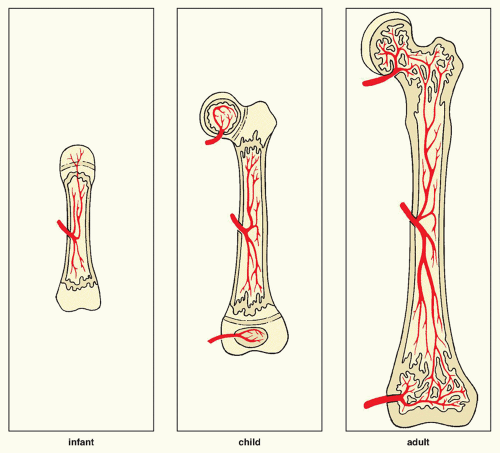
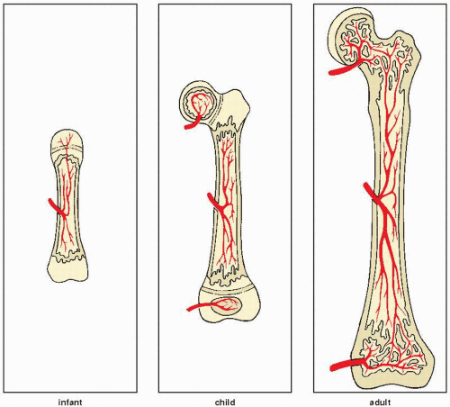
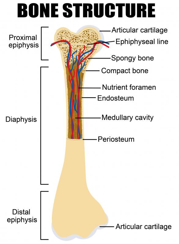
pediatric long bone anatomy
Giant Bone Island: Frontal and lateral knee radiographs show a large we have 15 Pictures about Giant Bone Island: Frontal and lateral knee radiographs show a large like Femur Anatomy and Attachments | Bone and Spine, Giant Bone Island: Frontal and lateral knee radiographs show a large and also How long does it take to heal a broken foot bone? | Tanglewood Foot. Here you go:
Giant Bone Island: Frontal And Lateral Knee Radiographs Show A Large
 www.pinterest.co.kr
bone island sclerotic lesion knee femur distal lateral well radiology spiculated giant circumscribed arrow ovoid frontal cortex margin xray radiographs
LONG BONE MEASUREMENT | Radiology Key
 radiologykey.com
bone measurement radiology
Anatomic Guidelines And Approaches For Biopsy Of The Long Bones
 radiologykey.com
approaches anatomic biopsy bones guidelines
How Long Does It Take To Heal A Broken Foot Bone? | Tanglewood Foot
 www.pinterest.com
broken foot fracture bones ray heal take bone does jones feet stress medical report know healing metatarsal cast care gardens
Solved: Identify The Bones And Features In Figures 15.7 And 15
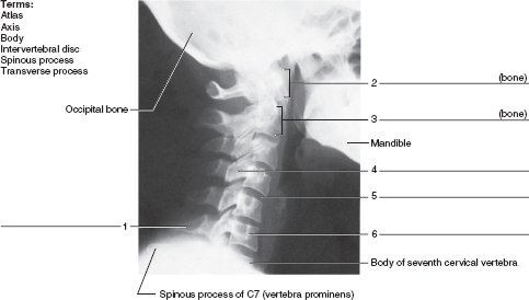 www.chegg.com
bones features identify radiograph neck bone label indicated lateral chegg figures provided solution chapter terms using
Radiologic Evaluation Of Musculoskeletal Infections | Radiology Key
 radiologykey.com
radiologic musculoskeletal evaluation infections bone
Osteology Unit - M.Y. Online Portfolio
 mkyousif17.weebly.com
lower bones anatomy skeletal system leg diagrams human diagram skeleton body bone labeled extremities osteology parts foot extremity upper tibia
Anatomy Of Bones In Childhood - Online Presentation
 en.ppt-online.org
anatomy
Bone Pictures - Labeled Drawn | Chandler Physical Therapy
 chandlerphysicaltherapy.net
labeled c2 c1 bone anatomy chandler physical therapy drawn frontier theme
Reconstruction Of Acetabular Bone Deficiencies Using The Antiprotrusio
 plasticsurgerykey.com
acetabular reconstruction cage bone surgery
Intramedullary Nailing Of Fractures | Bone And Spine
 boneandspine.com
intramedullary nailing nail femoral bone
Fracture Healing | Bone Healing, Fracture Healing, Orthopedic Nursing
 www.pinterest.com
bone healing fracture stages bones repair nursing stage remodeling does types fractured growth fractures orthopedic tissue which anatomy human xray
Radiologic Evaluation Of Musculoskeletal Infections | Radiology Key
 radiologykey.com
evaluation radiologic musculoskeletal infections bone vascular anatomy
Femur Anatomy And Attachments | Bone And Spine
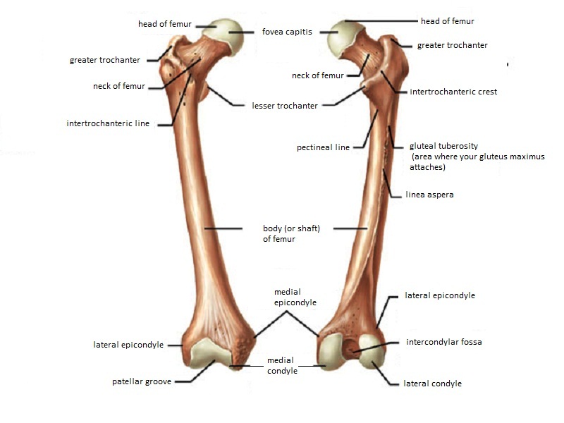 boneandspine.com
femur medially condyles convex forwards cylindrical widely
What Is The Haversian System? (with Pictures)
 www.wisegeek.com
haversian labeled shafts
Lower bones anatomy skeletal system leg diagrams human diagram skeleton body bone labeled extremities osteology parts foot extremity upper tibia. Radiologic musculoskeletal evaluation infections bone. Evaluation radiologic musculoskeletal infections bone vascular anatomy