knee anatomy bone and muscles
Knee MRI: sagittal PD | Image | Radiopaedia.org we have 8 Images about Knee MRI: sagittal PD | Image | Radiopaedia.org like The Knee | UT Health San Antonio, Lab 3 27 | Chandler Physical Therapy and also Art School Confidential: Summer Quarter 2012: Drawing & Anatomy: Bone. Here it is:
Knee MRI: Sagittal PD | Image | Radiopaedia.org
 radiopaedia.org
mri knee sagittal pd radiopaedia radiology case
Anatomy Of The Knee: An Introduction To The ACL - CHI Health Better You
 blogs.chihealth.com
anatomy
{Knee Joint Showing Image Of The Meniscus Along With The Cruciat
 www.johnthebodyman.com
knee joint ligaments meniscus posterior inferior along showing anatomy previous bones
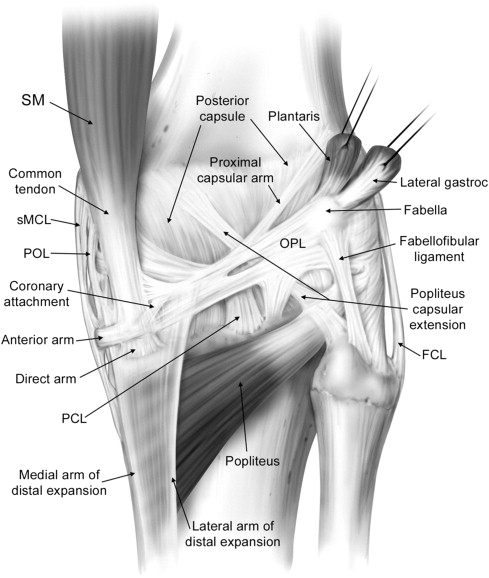
Posterolateral And Posteromedial Corner Injuries Of The Knee
 radiologykey.com
posterior posterolateral posteromedial mri injuries radiology laprade
Art School Confidential: Summer Quarter 2012: Drawing & Anatomy: Bone
 secondtryart.blogspot.com
drawing anatomy pelvis femur confidential bone
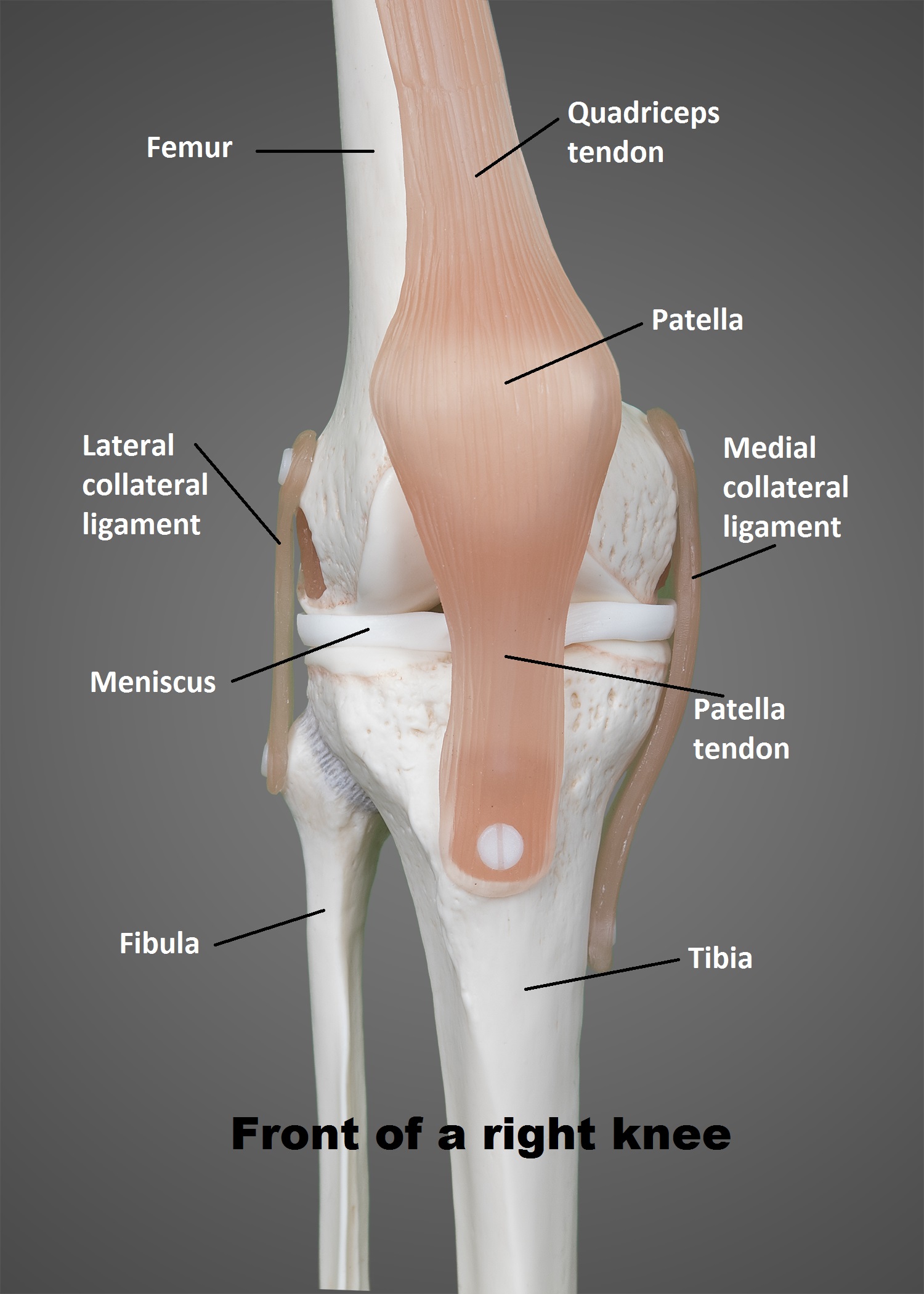
The Knee | UT Health San Antonio
 www.uthscsa.edu
anatomy physioactive basic ut physicians tissue
Anatomy Of The Patellar Tendon - Everything You Need To Know - Dr
 www.youtube.com
patellar tendon anatomy
Lab 3 27 | Chandler Physical Therapy
 chandlerphysicaltherapy.net
knee patella pain chondromalacia lower cap anatomy patellar labeled lab treatment tendonitis leg quadriceps muscle muscles joint chiropractor tibia az
Knee joint ligaments meniscus posterior inferior along showing anatomy previous bones. Knee mri: sagittal pd. {knee joint showing image of the meniscus along with the cruciat